INTRODUCTION
Solar observation and eye damage is known for millennia. Galileo was injured by telescopic solar observation 1. Solar retinopathy had been described in sailor, sunbather, military lookouts, photographer, religious fanatics and people under the influence of hallucinogenic drugs such as LSD 2. Solar eclipse is a geophysical phenomenon. It takes place when the moon appears between the sun and Earth. Periodically both the sun and moon return to the same position relative to one of the nodes, with the result, that eclipses occur at regular intervals. The time of the interval is called “Saros” and is about 6585.3 days or 18 years 9–11 days and 8 hours depending upon the intervening leap year. During one Saros, 70 eclipses takes place; 29 lunar, 41 solar. Of the later 10 are total while 31 are partial. During the 20th century, 375 eclipses occurred; 228 solar, 147 lunar. The last total eclipse was seen in USA on 11th July 1991. In Pakistan, India and certain parts of South Asia it was on 24th October 1995. Next total eclipse in USA will occur in 2017. Fifty million Mexicans watched total solar eclipse on 11th July 1991. Only 20 moderate cases were reported, all recovered after 4 months 3. In Mexico this was possible due to mass media awareness, epidemiological surveillance, early detection and use of sun filters.
The pathophysiology of Solar Eclipse Retinopathy (SE Retinopathy) is due to Retinal damage. The sun casts image on the retina, which is 160 μ in diameter. With 3mm pupil there is 4º rise in temperature whereas with 7 mm pupil there is 22º rise in temperature. During solar eclipse there is pupillary dilation. There is a variation in susceptibility to solar retinopathy, some individuals are relatively resistant and others develop symptoms after as little as 30–60 seconds exposure 4. Solar observation more than 90 seconds exceeds threshold of retinal damage 5. Brief solar observation during a solar eclipse is potentially dangerous because of the pupillary dilation and significant rise in temperature causing macular damage 6. People who have under gone cataract surgery or with retinal dystrophy or albinism or those taking photosensitizing medications are also at higher risk 7. Typical Eclipse retinopathy presents clinically as a small yellow white foveolar lesion. Over a period of 2–4 weeks lesion fades often to be replaced by foveolar depression or lamellar hole that produces reddish pit like reflex 8.
We carried out this study to observe the visual acuity changes in patients of solar eclipse retinopathy and their prognosis.
MATERIAL AND METHODS
This study was carried out in the department of Ophthalmology of Ayub Medical College following Solar Eclipse of April 29, 1995 for a seven years period up to April 2002. A total of 36 patients (45 eyes) were seen and included in the study. Diagnosis was based on history of voluntary solar eclipse exposure, poor visual acuity, after images, erythopsia and central scotoma.
Visual Acuity (VA) was recorded on Snellen chart. Slit lamp examination was done on all cases. Intraocular pressure (IOP) was measured with Goldman tonometer except in children. Fundoscopy was done and findings were recorded. Fundus photography and Fundus Fluorescein Angiography was done in patients with visual acuity of 6/60 due to limitation of resources. Twenty-one patients where visual acuity was 6/18 or less received oral steroids Methyl prednisolone 1–2 mg/Kg/day 3, topical non-steroidal anti-inflammatory drugs Diclofenac Sodium 0.1% Ocufen® / Optfen® TDS and topical steroids Prednisolone acetate 1% (Predforte®/ Optopred®) TDS. Patient with VA of 6/12 or more were given topical steroid and oral non-steroidal anti-inflammatory drugs Diclofenac sodium 75–150 mg daily. The patients were followed initially at weekly interval later at monthly interval and later on annually.
RESULTS
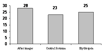
Blurred or poor vision was the most common symptoms and it appeared within 24 hours of exposure. After images were seen in 28 (77%) of the patients. Central scotoma was seen in 23 (63%) of the patients, whereas erythopsia was noted in 25 (69%) of the patients.
Figure 1: Presenting complaints in patients.
In 27 patients (75% of the cases) eye involvement was unilateral whereas 9 patients (25%) had bilateral involvement. Twenty-nine patients (80%) were male whereas 7 (19%) were female.
Table1: The Pattern of Eye Involvement in Patients.
Pattern of eye involvement |
No. |
Percent |
|
Unilateral |
27 |
75% |
|
Bilateral |
9 |
25% |
Age-wise analysis showed age group between 11 and 30 years were mostly affected in both sexes.
Table 2: Distribution of subjects by age
|
Age Group |
Male |
Female |
|
1-10 yrs |
3 |
1 |
|
11-20 yrs |
10 |
4 |
|
21-30 yrs |
7 |
2 |
|
31-40 yrs |
4 |
1 |
|
41-50 yrs |
2 |
1 |
|
51-60 yrs |
1 |
Nil |
|
61 and above |
Nil |
Nil |
Visual Acuity: Seven patients had VA of 6/9, 8 had VA of 6/12, 8 (22%) had VA of 6/18, 6 had VA of 6/24, 4 had VA of 6/36 while 3 patients presented with VA of 6/60.
Table 3: Presentation of visual acuity of patients
VA |
Number |
Percent |
|
6/60 |
3 |
8 |
|
6/36 |
4 |
11 |
|
6/24 |
6 |
16 |
|
6/18 |
8 |
22 |
|
6/12 |
8 |
22 |
|
6/9 |
7 |
19 |
A typical presentation was that Fundoscopy showed a small yellowish gray lesion in the center of foveolar area. Over the next two weeks this lesion gradually faded and was replaced by a small lamellar hole that produced a reddish pit like reflex adjacent to foveal reflex. This remained so during the follow up period and we considered it pathognomonic of SE Retinopathy, although a similar morphological appearance has been reported in ocular contusion and whip-lash injury 9.
Fluorescein angiography was done in three patients with VA of 6/60. It showed minor to moderate sub-foveal retinal pigment epithelium (RPE) transmission defect corresponding to lamellar hole.
Nine patients (25%) presented within first day of eclipse exposure, 16 patients (44.4%) after 2 days, 8 patients (22%) after 1 week, and 2 patients after 2 weeks. Only 1 patient came after 3 weeks. Patients who presented within first week and were started on treatment had good recovery. Patients who consulted after 2 weeks had poor recovery.
Table-4: Presentation of patients after exposure of eclipse.
|
Time of presentation |
Male |
Female |
|
After 1 day |
8 |
1 |
|
After 2 days |
10 |
6 |
|
After 1 week |
6 |
2 |
|
After 2 weeks |
2 |
Nil |
|
After 3 weeks |
1 |
Nil |
Complete recovery was noticed in 26 patients that occurred during the first 6 months where their VA returned to 6/6. Partial recovery where VA had improved to 6/12 or more was seen in 7 cases. No recovery was seen in 3 cases where the visual acuity remained 6/36 or below.
Table-5: Recovery of patients after treatment
|
Recovery |
No. |
VA |
|
Complete recovery |
26 |
6/6 |
|
Partial recovery |
7 |
6/12 or more |
|
No recovery |
3 |
6/36 or less |
DISCUSSION
Not all bands of electromagnetic radiations (EMR) emanating from the sun are in the visible spectrum and many of the non-visible bands can have a serious impact on biological functions although most harmful solar radiations are filtered out by atmosphere. The sunlight that reaches the earth contains sufficient amount of UV radiations to cause damage 2. Light can damage the retina by thermo-acoustic means as with Nd-Yag laser treatment, by thermal affects or burn as with xenon arc photo-coagulation or by photochemical mechanism as in solar retinitis or solar eclipse retinopathy 3.
The process is essentially similar to that outlined for lens although retina has more active metabolism with much higher O2 consumption and higher level of protection 10. Further more in retina, damaged protein can be protected by normal, cellular repair mechanisms. Short, intense, exposure to sunlight particularly with dilated pupil more than 90 seconds causes photochemical damage to photo-receptors. Similar damage could be caused by ophthalmic instruments like operating microscope 10.
Three patients with extrafoveal malignant melanoma volunteered to stare at the sun for one hour prior to enucleation. The eyes enucleated 36–48 hours post exposure showed varying degree of damage to RPE with oedema, irregular pigmentation and frank necrosis. In all three patients, VA returned to pre-exposure level prior to enucleation 11.
In experimental animals, rhesus monkey trained to identify the orientation of Landolt ring. Their VA returned to 6/6 in 30 days with blue light exposure, twice the threshold level of minimal lesion12. In two published reports approximately 50% of all the patients with SE Retinopathy returned to 6/6 vision within 6 months 13. The visual acuity showed better recovery and prognosis if patients were started on topical and oral steroids 14, 15. There is only one study which has followed the patient for fifteen years and reported better visual improvement in patients who presented early 16. In our study; complete recovery was noticed in 26 cases (72%). Recovery started after two weeks and no improvement was noticed after six months. In 10 patients (28%) the visual recovery was incomplete.
Conclusion
This is the only study about the prognosis of visual acuity in SE Retinopathy in Pakistan. Majority of the people knew that viewing a solar eclipse could be harmful to their eye but did not know the safest way17. We need to increase the public awareness regarding the safest method to watch an eclipse, which is by indirect method using projection. The higher number of patients presenting in our unit was due to lack of public awareness. Patients who presented early and were started on treatment had good recovery. Patients who presented after two weeks and had poor visual acuity at the time of presentation had poor prognosis. There are going to be 228 solar eclipses during this century. A national program has to be evolved to enhance mass awareness as that of Mexican model.
REFERENCES
1. The world book encyclopaedia. Volume 6 pp 35-37 World Book Inc 1985.
2. Cameron LL, Auer CL, McCormick PA. Association of sunlight with senile macular and lens changes. Invest Ophthalmol Vis Sci 1983; 24: 202.
3. Hochheimer BF, D’Anna SA, Calkins JL. Retinal damage from light. Am J Ophthalmol 1979;88:1039.
4. Gladstone GJ, Tasman W. Solar retinitis after minimal exposure. Arch Ophthalmol 1984;102:1510-12.
5. Ewald RA, Ritchey CL. Sun gazing as the cause of foveomacular retinitis. Am J Ophthalmol 1970;70:491.
6. Keightyley S. Solar retinopathy. Royal College of Ophthalmologists 1999;10:2-4.
7. Donne J. Staying safe during the eclipse. BMJ 1999;319:329-330.
8. Kuwabara T, Gom RA. Retinal damage by visible light: an electron microscope study. Am J Ophthalmol 1968;79:69.
9. Abebe MT, De Laey JJ. Foveo-macular retinitis as result of ocular contusion: Bull Soc Belge Ophthalmol 1992;243:171-5.
10. Penner R, Mc Nair JN. Eclipse blindness Am J Ophthalmol 1966; 61:1452.
11. Calkins JL, Hochheimer BF. Retinal light exposure from operating microscope. Arch Ophthalmol 1979; 97:2363.
12. Moon ME, Clarke AM, Ruffolo JJ Jr., Mueller HA, Ham, WT Jr. Visual performance in the rhesus monkey after exposure to blue light Vision Res 1978;18:1573.
13. Tso MOM, Lapiana FG. The human fovea after sun gazing. Trans AM Acad Ophthalmol 1975;79: 788.
14. Hatfield EM. Eye injuries and the solar eclipse: Sight Saving Rev. 1970;40:79.
15. Rosner M, Lam TT, Tso MO. Therapeutic parameters of methylprednisolone treatment for retinal photic injury in rat model. Res Commun Chem Pathol Pharmacol 1992;299-311.
15. Atmaca LS, Idil A, Can D. Early and late visual prognosis in solar retinopathy. Graefes Arch Clin Exp Ophthalmol 1995;233:801-4.
16. Nilofer SA, Badar SA, Iqbal SA. Assessment of knowledge, beliefs and practices of our population regarding effects of viewing a solar eclipse. Pak J Med Sci 2002;18(2): 117-121.