Difference in the outcome of patients managed with isolated
renal injury and co-existent abdominal organ injury
M. Amjad
Noor, M. Hammad Ather
The
Aga Khan University, Karachi.
Objective:
Involvement of associated intra-abdominal organs like
spleen; pancreas, bowel and liver with renal injuries have a
higher rate of open operative management. This is often done
to avert the potential of peri-renal infection and
subsequent risk of secondary hemorrhage of the injured
kidney after intra-abdominal surgery. With this background
we reviewed our experience to see if operative intervention
for co-existing injuries to intra-abdominal organs increase
the rate of nephrectomy for grade II-IV renal injuries.
Methods: In the period between January 1990 and December
2000, we identified 50 patients managed in this hospital
with evidence of external injury resulting in renal trauma.
Patients were divided into two groups; i) Patients with
isolated renal injury (group A) and ii) renal injury
associated with solid abdominal organ injury (group B). The
two groups were compared. The severity of renal injury was
classified by using the renal injury scale (I-V), which was
published by the Organ Injury Scaling (O.I.S.) Committee of
the American Association for the Surgery of Trauma (A.A.S.T.)
in 19891. Results: Sixty percent
patients had associated organ involvement. Penetrating
injuries were responsible for 47% patients in-group B
compared to only 5% in group A (p<0.001). CT was the
predominant radiological investigation in both groups.
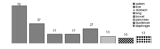
Spleen was the commonest intra-abdominal organ involved
(70%). Mean grade of injury in group-A was 2.2 compared to
2.7 in group B. Operative management was done in 20%
patients in group A compared to 29% in group B. Nephrectomy
in both groups were performed only for grade V injuries.
Conclusions: Exploration does not increase the rate of
nephrectomy; in group B grade II-IV injuries when explored
were all reconstructed. Penetrating injuries are more likely
to cause associated organ injuries (p<0.001). Spleen is the
commonest organ involved.
Key words:
kidney, trauma, intra-abdominal organ,
conservative, nephrectomy
Introduction
Advances in the imaging techniques have
not only made evaluation of patients with renal trauma
easier but has also impacted positively on the management.
Majority of hemodynamically stable patients, with grade I-IV
injuries are now managed conservatively1. In a
recent review of experience in the management of blunt renal
trauma, Danuser et al2 noted significant decrease
in the operative management in the period of 1989-1995
compared to 1973-1988.
The only absolute indication now for
renal exploration is persistent bleeding; other relative
indications include presence of non-viable tissue, urinary
extravasations, incomplete staging and arterial thrombosis.
In the absence of above factors, associated organ injury is
a relative indication for operative management. Involvement
of associated intra-abdominal organs like spleen; pancreas,
bowel and liver with renal injuries have a higher rate of
open operative management. This is often done to avert the
potential of peri-renal infection3 and subsequent
risk of secondary hemorrhage4 of the injured
kidney after intra-abdominal surgery. Hussmann3
et al noted 100% incidence of peri-nephric infections in
patients with co-existing colonic and/or pancreatic injuries
managed expectantly. With this in mind, we reviewed our
results in the last ten years in managing renal
trauma. With this background we
reviewed our experience to see if operative intervention for
co-existing injuries to intra-abdominal organs increase the
rate of nephrectomy for grade II-IV renal injuries.
Methods
In the period between January 1990 and December 2000, we
identified 50 patients managed in this hospital with
evidence of external injury resulting in renal trauma.
Patients were identified using ICD-9cm5.
The relationships of
genitourinary injury with associated organ injuries, age and
gender of the patient, the mechanism of injury, the mode of
treatment, the mortality and morbidity were evaluated.
Patients presenting to the emergency room with general
abdominal trauma, and diagnosed and then hospitalised for
renal trauma were included in the study. After initial
clinical evaluation with history and physical examination,
routine blood count, renal function test and urinalyses were
preformed on each patient. All patients were subsequently
evaluated radiologically using ultrasound, intravenous
urogram (IVU) and/or CT Scan. Ultrasound was performed
either as a screening test in vitally stable patients with
no or minimal microscopic haematuria (2–5 erythrocytes/High
Power Field) or as tool for continuous evaluation in
patients managed conservatively. IVU was performed either as
an emergency one shot IVU prior to laparotomy and after
resuscitation and haemodynamic stabilization (systolic blood
pressure of ³90 mm
Hg.) or in haemodynamically stable patients with haematuria
but no clinical evidence of associated intra-abdominal organ
involvement.
Patients were divided into
two groups; i) Patients with isolated renal injury (group
A), and ii) renal injury associated with solid abdominal
organ injury (group B). There were 20 patients in Group A
and 30 belonged to Group B. The two groups were compared.
The severity of renal injury was classified by using the
renal injury scale, which was published by the Organ Injury
Scaling (OIS) Committee of the American Association for the
Surgery of Trauma (AAST) in 19891. Statistical
analysis was performed on a commercially available software
i.e. SPSS (statistical package for social sciences).
Results
In the period between January 1990 and
December 2000, 50 patients were identified. There were 20
patients in Group A and 30 belonged to Group B. Gender
distribution between the two groups was similar (9:1 male to
female). In-group A penetrating injuries accounted for only
5% of the patients whereas 95% had blunt abdominal trauma
whereas in Group B 47% patient had penetrating injuries
compared to 53% blunt abdominal injury. All patients in both
groups had microscopic or gross haematuria. . In Group A 45%
patients had a CT scan and the rest had ultrasound and/or
IVU. In Group A 45% patients had
a CT scan and the rest had ultrasound and / or IVU. In group
B 3/4th of the patients were evaluated by CT scan and 1/4th
by ultrasound and / or IVU.
Details of associated organ involvement
are shown in Figure-1.
Mean grade of injury for Group A was 2.2±0.3
compared to Group B, which were 2.7±0.25
and is shown in Figure-2.
One fifth of the patients in group A and
about a third in group B were managed by open surgery, the
details of which are described in table-1.
Table-1: Intervention in relation to the grade of injury.
|
Grade (n) |
Isolated Renal Injury
n=20 |
Associated Organ Injury
n=30 |
|
Operative |
Conservative |
Operative |
Conservative |
|
I (11) |
- |
7 |
- |
4 |
|
II (18) |
- |
5 |
1 |
12 |
|
III (10) |
- |
5 |
1 |
4 |
|
IV (5) |
1 |
2 |
2 |
- |
|
V (6) |
1 |
1 |
5 |
- |
Complications were noted in 15% in group
A and 16 % in group B. In group A one patient had
significant bleeding requiring blood transfusions. One
patient developed new onset hypertension and one patient
developed a urinary fistula, which responded to placement of
ureteric stent. In group B one patient each had significant
bleeding, urinary fistula and a perinephric collection
requiring percutaneous drainage whereas two patients
developed new onset hypertension. In group A there was no
mortality whereas in group B 10% died. The details of which
are described in Table-2.
Table-2: Mortalities in Group B.
|
Age, Gender |
Mechanism
|
Grade
|
Operative procedure |
Cause of death |
|
25 yrs, Male |
Gunshot |
Grade 5 |
Thoracotomy, spleenectomy, distal
pancreastectomy, left nephrectomy |
Cardiac arrest |
|
32 yrs, Male |
Gunshot |
Grade 5 |
Hepatic laceration and duodenal
perforation repair, exteriorization of
gastric antrum, Right nephrectomy |
Hypovolumic shock |
|
48 yrs, Male |
Gunshot |
Grade 2 |
Spleenectomy, loop colostomy,
laminectomy |
Anterolateral
Myocardial Infarction,
Aspiration pneumonia |
Among four operative interventions in
Group A one patient had a partial nephrectomy, another had a
repair of parenchymal laceration and two patients had a
nephrectomy. In Group B however, four patients had repair of
laceration and drainage of haematoma while 2 had partial
nephrectomy and three patients had a nephrectomy done
(Figure-3).
Discussion
Trauma is a leading cause of death in
young population4. However, deaths related to
renal injuries alone are rare. Mortality and morbidities are
seen in patients with associated intra-abdominal organ
injuries, and cerebral, thoracic and skeletal injuries. It
is therefore important to assess the presence of related
organs involved.
In the recent years with advances in
radiological evaluation and increasing experience in the
management vast majority of patients with renal trauma are
managed conservatively. In the experience of McAninch et al6
the frequency, of accompanying intra-abdominal organ
injuries was 80%. In the present study incidence of
accompanying intra-abdominal organ injuries was 60%. The
most commonly involved organs were spleen followed by liver
and bowel (Table-1).
All patients with injuries to the solid organs of the
abdomen and who are haemodynamically stable should be
considered candidates for non-operative management after
their injuries have been staged by abdominal CT scanning.
One limitation of CT in the presence of intra-abdominal
organ injury is that the stage of the injury determined does
not always predict which patients would require laparotomy7.
These patients require close haemodynamic monitoring for
early recognition of an associated hollow viscus injury in
need of repair, if the non-operative approach fails.
Although delayed bleeding from the liver seems extremely
rare, delayed rupture of the spleen and continued
haemorrhage into the retro peritoneum from an injured kidney
are not unusual, so patients with spleenic and renal
injuries should be considered candidates for repeat imaging
procedures before discharge. Others likely to benefit from a
second look at their injuries include patients with sub
capsular haematomas, patients with recognized extravasations
on the initial scan, and athletes anxious to return to
contact sports.
Experience from major trauma centres suggests that the
incidence of missed intestinal injuries is low in adults and
children managed non-operatively, but surgeons must be
diligent in monitoring for increasing abdominal pain,
abdominal distension, vomiting, and signs of inflammation,
which may be delayed manifestations of intestinal
disruption.
Patients with vascular injuries (grade V
injuries to the spleen, liver, or kidney) may be candidates
for interventional imaging procedure, such as
angio-embolization or stenting, but some of these patients
are best served by immediate laparotomy7.
Patients with associated intra-abdominal
injuries, however, often require laparotomy, which provides
an opportunity to repair major renal lacerations and drain
haematoma. The concern with this approach is that it may
increase the rate of nephrectomy performed as an expedient
procedure to attain haemodynamic stability8. On
the other hand, non-operative treatment in patients with
pancreatic and colonic injuries places them at significant
risk for associated urologic complications. Particularly
patients who have colonic injuries are at an increased risk
of septic complication with devitalizing renal injuries3.
Once the abdomen is opened and intra-abdominal injuries are
tackled in a stable patient, renal pedicle is isolated
before exploring the kidney. This maneuver maximizes renal
preservation and reconstruction of damaged kidney with
minimal blood loss. Utilizing this approach McAninch et al9
were able to achieve 88% renal salvage with minimal
morbidity. However, proper patient selection is important as
majority of patients with grade
II and
III injuries and
no evidence of devitalized renal parenchyma and absence of
pancreatic and colonic injuries could still be managed
conservatively.
References
1.
Moore EE, Shackford SR, Pachter HL,
McAninch JW, Browner BD, Champion HR, Flint LM, Gennarelli
TA, Malangoni MA, Ramenofsky ML, et-al. Organ injury
scaling: spleen, liver, and kidney. J Trauma. 1989; 29:
1664-6
2.
Danuser H, Wille S, Zoscher G, Studer
U E. How to treat blunt kidney rupture: Primary open surgery
or conservative treatment with deferred surgery when
necessary? Eur Urol 2001; 39:9-14
3.
Husmann DA, Gilling PJ, Perry MO,
Morris JS, Boone TB. Major renal lacerations with a
devitalized fragment following blunt abdominal trauma: a
comparison between non-operative (expectant) versus surgical
management [see comments] J-Urol. 1993; 150: 1774-7
4.
Rosenberg ML, Fenely MA. Conference on injury in
America: a summary. Public health Rep. 1987; 102:577
5.
International classification of
Disease, 9th edition- clinical modification. Hart
AC, Hopkins CA (eds). Janetshack St Anthony
publications, Salt lake city, Utah: 2002
6.
Miller KS, McAninch JW. Radiographic
assessment of renal trauma: our 15-year experience. J Urol.
1995; 154: 352-5
7.
Knudson MM, Maull KI. Non-operative
management of solid organ injuries. Past, present, and
future. Surg Clin North Am. 1999; 79: 1357-71
8.
Husmann DA, Morris JS. Attempted
non-operative management of blunt renal lacerations
extending through the cortico-medullary junction: the
short-term and long-term sequelae [see comments] J Urol.
1990; 143: 682-4
9.
McAninch JW, Carroll PR, Klosterman
PW, Dixon CM, Greenblatt MN. Renal reconstruction
after injury. J Urol. 199; 145: 932-7